Big New Genome Wide Association Study On Androgenetic Alopecia - Preprint

Armando Jose

Senior Member
My Regimen
Reaction score
975
"At the start of the growth phase (anagen), cells at the base of the bulge (hair germ, HG), which are initially similar to bulge HF-SCs in gene expression (Greco et al., 2009), become proliferative, grow downward, and engulf the transient mesenchymal niche component (dermal papilla, DP) as they transition to committed, so-called transit-amplifying matrix cells (TACs)."

https://www.ncbi.nlm.nih.gov/pmc/articles/PMC3856689/
 

InBeforeTheCure

Established Member
My Regimen
Reaction score
950
"LHX2 loss leads to a failure to maintain HF-SC quiescence and hair anchoring, as well as progressive transformation of the niche into a sebaceous gland."

So Olsen (2003) thinks that the hair follicle dies entirely. Cotsarelis showed us that the stem cells remain, but the progenitor were depleted and Fuchs et al think that over time the bulge becomes a sebaceous gland over time, which I suppose suggests that the stem cells dies and there's basically no coming back from that.

But the study by Fuchs was about Lhx2 knockout mice, not A.G.A...

At this stage I think we need to make a sticky dedicated to the actual architecture of the hair follicle.

A sticky for resources on hair follicle structure, morphogenesis, cycling, biochemistry and so on is a great idea. :)
 

Gone

Established Member
My Regimen
Reaction score
96
But the study by Fuchs was about Lhx2 knockout mice, not Androgenetic Alopecia...

In your post you said that Lhx2 was found by Cotseralis to be downregulated in bald scalp. Bald human scalp? Or mouse, (I know now that the Fuchs one is referring to mice, I'm referring to Cotseralis's.)
 

InBeforeTheCure

Established Member
My Regimen
Reaction score
950
In your post you said that Lhx2 was found by Cotseralis to be downregulated in bald scalp. Bald human scalp? Or mouse, (I know now that the Fuchs one is referring to mice, I'm referring to Cotseralis's.)

Bald human scalp, guys with A.G.A., yeah.
 

Beowulf

Established Member
My Regimen
Reaction score
129
A sticky for resources on hair follicle structure, morphogenesis, cycling, biochemistry and so on is a great idea. :)

I'm about 500 words in and I'm already super confused. I'm just going to try to write a draft and you guys can have a look and we'll see what we come up with.
 

Swoop

Senior Member
My Regimen
Reaction score
1,332
I was quoting IBTC, so Fuchs et al think that the bulge can become a sebaceous gland. Swoop a few months ago mentioned that Olsen said that the Dermal papilla dies and that's it. (https://books.google.com.au/books?id=pHrX2-huQCoC&pg=PA185&lpg=PA185&dq=olsen+2003+hair&source=bl&ots=4xsJjsuJjD&sig=rLTF9h-kIja8vTP8QMoOKzMxw64&hair loss=en&sa=X&ved=0ahUKEwjWlbqt8cTQAhXBvI8KHXKlBB0Q6AEIMzAI#v=onepage&q=olsen 2003 hair&f=false)

You mean this quote right?

"The end result is the production of smaller, unpigmented vellus hairs instead of larger, pigmented terminal hairs. Although the follicles do progressively get smaller, the overall scalp follicle density appears to be preserved until late in the course of hair loss when follicles are eventually destroyed (for a thorough description, see Olsen, 2003)."

I can't open your link though.. I want to read that from Olsen. Can anyone get it?
 

Swoop

Senior Member
My Regimen
Reaction score
1,332
reversible.jpg
 

Armando Jose

Senior Member
My Regimen
Reaction score
975
Hair loss is not due at exhaustion of the follicular stem cells.

when the hair is lost, the system of stem cells must be maintained, ... a proof is that the epidermis continues to renew every month and little.

Also in this type of non-cicatricial alopecia what is generally observed is that the hair disappears or is miniaturized, and the attached sebaceous gland is maintained.

In muy opinions it correct
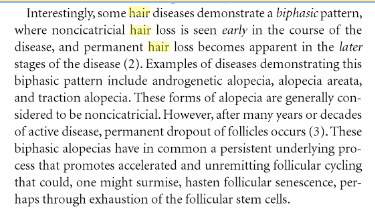
"Persistent underlying process that promotes accelerated and unremitiling follicular cycling, that could, one might surmise, hasten follicular senescence, perhaps trought exhaustion of the follicular stem cells."
 

Beowulf

Established Member
My Regimen
Reaction score
129
You mean this quote right?

"The end result is the production of smaller, unpigmented vellus hairs instead of larger, pigmented terminal hairs. Although the follicles do progressively get smaller, the overall scalp follicle density appears to be preserved until late in the course of hair loss when follicles are eventually destroyed (for a thorough description, see Olsen, 2003)."

I can't open your link though.. I want to read that from Olsen. Can anyone get it?

I believe so, it came with your awesome Tsuji gif. In any case I was too hasty follicle does not necessarily refer to dermal papilla.
 

InBeforeTheCure

Established Member
My Regimen
Reaction score
950
Ayy look at this. The exact function of the SNP (rs12203592) in Intron 4 of IRF4 associated with A.G.A. is already known. In melanocytes, this SNP affects binding affinity of TFAP2A to this region. IRF4 acts together with MITF to enhance transcription of tyrosinase, which is an enzyme involved in melanin synthesis. The T allele, which is associated with A.G.A. as well as with lighter pigmentation, disrupts binding of TFAP2A to Intron 4 of IRF4 and so less IRF4 is transcribed. Therefore, the melanocytes produce less melanin...and your chances of going bald increase???

Sequence polymorphisms linked to human diseases and phenotypes in genome-wide association studies often affect non-coding regions. A single nucleotide polymorphism (SNP) within an intron of the gene encoding Interferon Regulatory Factor 4 (IRF4), a transcription factor with no known role in melanocyte biology, is strongly associated with sensitivity of skin to sun exposure, freckles, blue eyes and brown hair color. Here we demonstrate that this SNP lies within an enhancer of IRF4 transcription in melanocytes. The allele associated with this pigmentation phenotype impairs binding of the TFAP2A transcription factor which together with the melanocyte master regulator MITF, regulates activity of the enhancer. Assays in zebrafish and mice reveal that IRF4 cooperates with MITF to activate expression of Tyrosinase (TYR), an essential enzyme in melanin synthesis. Our findings provide a clear example of a non-coding polymorphism that affects a phenotype by modulating a developmental gene regulatory network.

Source: A polymorphism in IRF4 affects human pigmentation through a tyrosinase-dependent MITF/TFAP2A pathway

So tell me...How exactly is less melanin synthesis contributing to A.G.A. pathogenesis? o_O
 

Gone

Established Member
My Regimen
Reaction score
96
Is it not possible that Androgenetic Alopecia inhibits melanin? Instead of the other way around. We know that PGF2a increases melanin and grows hair.
 

InBeforeTheCure

Established Member
My Regimen
Reaction score
950
Is it not possible that Androgenetic Alopecia inhibits melanin? Instead of the other way around. We know that PGF2a increases melanin and grows hair.

Not in this case, because if it was the other way around, why would this SNP show up in a GWAS?
 

Grasshüpfer

Experienced Member
My Regimen
Reaction score
636
Not in this case, because if it was the other way around, why would this SNP show up in a GWAS?

Fits perfectly in with Yae-olde-Vitamin-D-Androgenetic Alopecia-theory. Please shoot me down if i'm moving in S.Foote territory. :D

Melatonin is associated with Vitamin D production and low melatonin expession is an adjustment to the low light, low vitamin D conditions in eg. northern europe. Vitamin D deficency is associated with autoimmune reactions, so the low melanin could be an adjustment to an underlying issue.
"Deficiency in vitamin D is associated with increased autoimmunity as well as an increased susceptibility to infection."
https://www.ncbi.nlm.nih.gov/pmc/articles/PMC3166406/

The line of thinking works for AA, but how do autoimmune reactions fit in with Androgenetic Alopecia?

Autoimmune reactions are discussed to be involved in early artheriosclerosis.
Innate immune mechanisms have been demonstrated in atherosclerosis, particularly in early stages of the disease.
https://bmcmedicine.biomedcentral.com/articles/10.1186/1741-7015-12-47


And DHT is apparently supressing inflammation in hypoxic envionments.
In contrast, during hypoxia, DHT suppressed NF
kappa.gif
B activity and COX-2. Thus, androgens may have a differential impact on regulation of inflammation during pathophysiological compared to normal physiological conditions.
http://www.fasebj.org/cgi/content/meeting_abstract/21/6/A1385-a


This would explain why we see those insane dht concentrations in the scalp compared to serum. So Androgenetic Alopecia would be a side effect of the body sucessfully regulating inflammation in the scalp.


Well the theory is very very thin lol. Lets try to shoot it down, see if it has any validity.
 

InBeforeTheCure

Established Member
My Regimen
Reaction score
950
First of all, I went too far in assuming the important thing here must be melanin synthesis. IRF4 surely has target genes in melanocytes unrelated to melanin synthesis, and maybe something else produced in melanocytes is contributing to A.G.A. somehow. The two main possibilities are the following:

1) A failure to properly adjust for population stratification has resulted in a spurious association between rs12203592 and A.G.A. Since the T allele at rs12203592 is most common in Europeans and non-existent in Africans and East Asians, and there are immigrants in the UK who probably have a lower rate of A.G.A. than native Brits, if the adjustment for this isn't quite right you could see a meaningless correlation between the two. They do say they corrected for population stratification though (it's standard practice in GWAS for obvious reasons), so it would have to be something more subtle than a total failure to correct for it.

Statistical Analysis
A genome-wide association study was conducted using baldness pattern residuals as the
dependent variable. The residuals were obtained from a linear regression model of baldness
pattern on age, assessment centre, genotyping batch and array, and 10 principal components
to correct for population stratification.

OR...

2) Lower expression of IRF4 in melanocytes is a risk factor for A.G.A.

Fits perfectly in with Yae-olde-Vitamin-D-Androgenetic Alopecia-theory. Please shoot me down if i'm moving in S.Foote territory. :D

Dream big, Grasshupfer. If we develop these crazy ideas further, we could pass S. Foote and reach Ernie Primeau territory. ;)

Melatonin is associated with Vitamin D production and low melatonin expession is an adjustment to the low light, low vitamin D conditions in eg. northern europe. Vitamin D deficency is associated with autoimmune reactions, so the low melanin could be an adjustment to an underlying issue.
"Deficiency in vitamin D is associated with increased autoimmunity as well as an increased susceptibility to infection."
https://www.ncbi.nlm.nih.gov/pmc/articles/PMC3166406/

The line of thinking works for AA, but how do autoimmune reactions fit in with Androgenetic Alopecia?

Autoimmune reactions are discussed to be involved in early artheriosclerosis.
Innate immune mechanisms have been demonstrated in atherosclerosis, particularly in early stages of the disease.
https://bmcmedicine.biomedcentral.com/articles/10.1186/1741-7015-12-47


And DHT is apparently supressing inflammation in hypoxic envionments.
In contrast, during hypoxia, DHT suppressed NF
kappa.gif
B activity and COX-2. Thus, androgens may have a differential impact on regulation of inflammation during pathophysiological compared to normal physiological conditions.
http://www.fasebj.org/cgi/content/meeting_abstract/21/6/A1385-a


This would explain why we see those insane dht concentrations in the scalp compared to serum. So Androgenetic Alopecia would be a side effect of the body sucessfully regulating inflammation in the scalp.

But lighter pigmentation -> more efficient absorption of UV radiation, right?

The variant in IRF4 associated with increased A.G.A. risk leads to lighter skin and therefore more efficient absorption of UV radiation (which is probably why it was selected for in low-UV climates), and therefore higher vitamin D synthesis than would otherwise be the case.

Well the theory is very very thin lol. Lets try to shoot it down, see if it has any validity.

It has seemingly absurd consequences, like someone who lives in Los Angeles would be more likely to go bald than someone who lives in Seattle. Surely if that were the case we would have noticed by now, so in my mind this interpretation of the data fails from the start.

BTW, a new study on this very subject just so happened to come out a few days ago, although I haven't read it yet.

Abstract: Mice and human patients with impaired vitamin D receptor (VDR) signaling have normal developmental hair growth but display aberrant post-morphogenic hair cycle progression associated with alopecia. In addition, VDR–/– mice exhibit impaired cutaneous wound healing. We undertook experiments to determine whether the stress-inducible regulator of energy homeostasis, DNA damage-inducible transcript 4 (Ddit4), is involved in these processes. By analyzing hair cycle activation in vivo, we show that VDR−/− mice at day 14 exhibit increased Ddit4 expression within follicular stress compartments. At day 29, degenerating VDR−/− follicular keratinocytes, but not bulge stem cells, continue to exhibit an increase in Ddit4 expression. At day 47, when normal follicles and epidermis are quiescent and enriched for Ddit4, VDR−/− skin lacks Ddit4 expression. In a skin wound healing assay, the re-epithelialized epidermis in wildtype (WT) but not VDR−/− animals harbor a population of Ddit4- and Krt10-positive cells. Our study suggests that VDR regulates Ddit4 expression during epidermal homeostasis and the wound healing process, while elevated Ddit4 represents an early growth-arresting stress response within VDR−/− follicles.

Keywords: wound repair; VDR; mTOR; DDIT4; stress; hair follicle; stem cells
 

Grasshüpfer

Experienced Member
My Regimen
Reaction score
636
Found an article on a similar gene, which has an effect on perceived age.
  • Variants in MC1R, a pigmentation gene, significantly associated with perceived age
  • The MC1R association was independent of wrinkling, skin color, and sun exposure
http://www.cell.com/current-biology/fulltext/S0960-9822(16)30184-1

Individuals carrying the homozygote MC1R risk haplotype looked on average up to 2 years older than non-carriers. This association was independent of age, sex, skin color, and sun damage (wrinkling, pigmented spots) and persisted through different sun-exposure levels. Hence, a role for MC1R in youthful looks independent of its known melanin synthesis function is suggested.

Does mc1r show up in the Androgenetic Alopecia gwas?

Regardingy my awesome theory.. I think there is a link between microcirculation and the effect of certain hormones on hair.
But that's honestly a forking theory of this thread, so I might open an other one.
 

InBeforeTheCure

Established Member
My Regimen
Reaction score
950
Found an article on a similar gene, which has an effect on perceived age.
  • Variants in MC1R, a pigmentation gene, significantly associated with perceived age
  • The MC1R association was independent of wrinkling, skin color, and sun exposure
http://www.cell.com/current-biology/fulltext/S0960-9822(16)30184-1

Individuals carrying the homozygote MC1R risk haplotype looked on average up to 2 years older than non-carriers. This association was independent of age, sex, skin color, and sun damage (wrinkling, pigmented spots) and persisted through different sun-exposure levels. Hence, a role for MC1R in youthful looks independent of its known melanin synthesis function is suggested.

Does mc1r show up in the Androgenetic Alopecia gwas?

Look at Post #10 on pg. 1 of this thread -- I actually listed it there. ;) However, that region of Chr16 is extremely gene-dense, so there are a lot of possibilities.

From the paper you posted:

In conclusion, this study is the first to identify genetic variants
significantly associated with perceived age. We provide evidence
that, of eight million tested, DNA variants in the MC1R
gene had the strongest association with perceived age in subjects
of European ancestry, and a MC1R compound marker
was genome-wide significant independently of age, sex, skin
color, sun exposure, wrinkles, and pigmented spots. Follow-up
work on how the MC1R protein is affecting facial aging, for
example, through non-UV pro-oxidant phenomelanin effects
[22] or fibroblast function [21], is now required.
Moreover, as
this study demonstrates that a GWAS of perceived facial age
is indeed feasible, future studies using large consortia GWASs
should be performed to identify additional genetic loci that associate
with perceived facial age. Expectedly, this will provide
further insights into the biological pathways that underlie variation
in facial aging and eventually on the utility of genotypebased
prediction of perceived age alongside chronological age
estimation from molecular biomarkers.

Reference #22 refers to this paper, where they find pheomelanin (which is increased with lower MC1R expression, or with loss-of-function mutation such as that associated with red hair) increases oxidative damage.

To determine whether ROS-mediated oxidative DNA damage is
affected by the pheomelanin synthesis pathway, levels of 8,59-cyclo-29-
deoxyadenosine (cdA) and 8,59-cyclo-29-deoxyguanosine (cdG) were
measured inDNA isolated from skin of red-Mc1re/e and albino-Mc1re/e
mice, using a previously reported liquid chromatography-tandem
mass spectrometric method24 (Fig. 4c). These two ROS-mediated
cyclopurines are unlikely to be artificially induced during sample preparation
and are quite stable25,26. Significantly, replication studies in
Escherichia coli have shown that S-cdA and S-cdG can lead to A-to-T
and G-to-A mutations at frequencies of 11% and 20%, respectively27.
Comparing cyclopurine levels in the skin of various pigmentation variant
mice, it was found that the levels of cdA and cdG are significantly
higher in skin from red-Mc1re/e mice compared to skin from
albino-Mc1re/e animals (Fig. 4d, e). This observation indicates that
activation of the pheomelanin synthesis pathway results in increased
oxidative DNA damage. Correlative evidence for increased cellular
oxidative stress was also found in the observation that red-Mc1re/e
mouse skin carries higher levels of lipid peroxides, a product of
ROS-mediated lipid damage (Fig. 4f ).

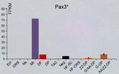
Besides IRF4 and perhaps MC1R, there's also PAX3 associated with A.G.A., which is a melanocyte signature gene according to Rendl's "Hair-GEL" site.

Pax3.png

Mc = melanocytes

A snippet from the IRF4 study:

Freckles represent clusters of concentrated melanin in the skin, without an increase in melanocyte numbers. Freckles are either light brown or red but usually become darker and more visible upon exposure to sunlight. GWAS studies have shown that variations in MC1R, ASIP, TYR, and BNC2, in addition to IRF4, are associated with freckles (Sulem et al., 2007; Sulem et al., 2008; Eriksson et al., 2010). Mutations in the MC1R gene have been highly associated with the formation of freckles (Bastiaens et al., 2001). In addition to regulating TYR and IRF4 gene expression, MITF is also known to regulate expression of MC1R (Adachi et al., 2000; Smith et al., 2001; Aoki and Moro, 2002). This suggests the possibility that, like with TYR, IRF4 cooperates with MITF in regulating expression of MC1R in melanocytes. Indeed, ChIP-seq studies show that MITF binds MC1R regulatory sequences (Strub et al., 2011).

The effects of both TFAP2A and IRF4 are mediated through MITF which is needed both for the TFAP2A-mediated activation of IRF4 and the IRF4-mediated activation of TYR. ChIP-seq studies suggest that MITF regulates TFAP2A as well as IRF4 (Strub et al., 2011). The cooperative effects of MITF and IRF4 suggest that these proteins might physically interact on the TYR promoter. TFAP2A is expressed in a variety of both mesenchymal and epithelial cell types. It is not known how MITF and TFAP2A interact in the regulation of IRF4. Since the binding sites are physically close to each other, it is possible that both proteins are part of a larger complex formed at the site. Together, these data suggest that transcription factors which are expressed in multiple cell types can form cell-type specific complexes to govern differentiation.

So we could postulate that GWAS hits PAX3, IRF4, TFAP2A, MC1R, ASIP, and C10orf11 (which is required for melanocyte differentiation) could be part of a melanocyte subnetwork which is somehow involved in A.G.A., with increased ROS production being a possible factor, but there are probably other possibilities.
 
Last edited:

Grasshüpfer

Experienced Member
My Regimen
Reaction score
636
Actually i checked for mc1r myself after my post but then fell asleep. :D

I just looked at the abstract of the gwas again. I wonder how much the prediction accuracy changes if you take just the top 20 associated hits instead of all 250. 250 is so fuzzy still.

pfffff... I know just enough about molecular biology that im aware of my limitations. I am still treating it like the code we write for computers while we already know that it is at least one order of magnitude more complex through epigenetic influences.

Feels so much better to go back to really dumb theories. :D
- "Why are you looking for your keys under the street lamp, you lost them over there in the bushes, didn't you?"
- "Yes, but here at least i can see something."
 

Armando Jose

Senior Member
My Regimen
Reaction score
975
- "Why are you looking for your keys under the street lamp, you lost them over there in the bushes, didn't you?"
- "Yes, but here at least i can see something."

+1

The key question is why common hair loss only affects certains hairs?
 

InBeforeTheCure

Established Member
My Regimen
Reaction score
950
I just looked at the abstract of the gwas again. I wonder how much the prediction accuracy changes if you take just the top 20 associated hits instead of all 250. 250 is so fuzzy still.

It's more like 58 regions, 56 autosomal and 2 X-chromosome, rather than 250. Those 250 include "windows" which are right next to each other and probably influence the same gene.

+1

The key question is why common hair loss only affects certains hairs?

That's a complicated question which has barely been explored, so it will probably be many years before we have a satisfactory answer to it. I think an even more interesting question is why susceptibility to androgen-induced hair loss "switches on" in particular HFs at a particular age. Hamilton showed that while 20-year-old castrates injected with testosterone lose hair at the same rate as normal 20-year-olds, but castrates injected with testosterone in their 60s can lose all of their hair within months. It seems then that in these 60-year-old castrates, there is some latent androgen sensitivity which exists independent of exposure to androgens, but it needs that DHT trigger to set it off.
 

Armando Jose

Senior Member
My Regimen
Reaction score
975
Hamilton studies are unique, in the 40', and not replicated. With a grain of salt, if you read them....
it is not a dogma, in my opinion
 
Top