Successful Treatment of Alopecia Areata with Topical Vitamin D

Jacob

Senior Member
Reaction score
44
Dovonex?

I wouldn't get too excited about the korean kiddo. Again..different alopecia...short hairloss period- " 2-month history of sudden hair loss on the vertex region of the scalp"..etc. But then again..they must have other examples out there somewhere...right?
 

squeegee

Banned
Reaction score
132
I think that the damaged receptors are just symptom of ROS/inflammation which comes with hairloss.. The kid doesn't have male pattern baldness.. no DHT is involved yet..
 

resu

Senior Member
Reaction score
1,334
Yes there's been some noise regarding damaged d3 receptors, I should start saving all the info I read. It's an angle that hasn't been tried, who could forget all the buzz from last year regarding vitamin d and hair loss (not supplements related).
 

squeegee

Banned
Reaction score
132
Vitamin D receptor depletion by Wnt/beta-catenin signaling caused alopecia areata in human

Vitamin D receptor (VDR) is expressed in the epidermal component of hair follicle and dermal papilla cell and the lack of VDR results alopecia. In this study, we investigated the VDR expression in the human normal follicle and the alopecia areata (AA). First, we demonstrated human tissue was taken for immunohistochemical staing for β-catenin, Wnt3a, Wnt5a, Wnt10b, proliferating cell nuclear antigen (PCNA), involucrin and filaggrin.
Our results showed that VDR expressed in the epidermal keratinocytes of normal follicle, but reduced in the AA. We also found that β-catenin, Wnt3a, Wnt5a and Wnt10b, all of which are associated with hair growth, these strong observed in normal follicle, whereas showed decreased expression in AA. To investigate the relationship between VDR depletion and epidermal differentiation, we examined PCNA, involucrin and filaggrin in the hair follicle and epidermis of scalp. Epidermal differentiation markers, involucrin and filaggrin, showed reduced expression levels in AA, and PCNA were less induced in the hair follicle of AA.
DKK1, a canonical Wnt inhibitor, which were involved VDR and β-catenin. In order to determine affected by dickkopf 1 (DKK1) using human primary keratinocyte, Western blot analysis was performed. DKK1 treatments reduced the VDR levels, indicating that the VDR exhaustion results from inhibited Wnt signaling and lead to epidermal differentiation inhibition.
In summary, we showed that VDR depletion by Wnt/β-catenin signaling induce AA in human. These results suggest the potential use of AA therapy.

- - - Updated - - -

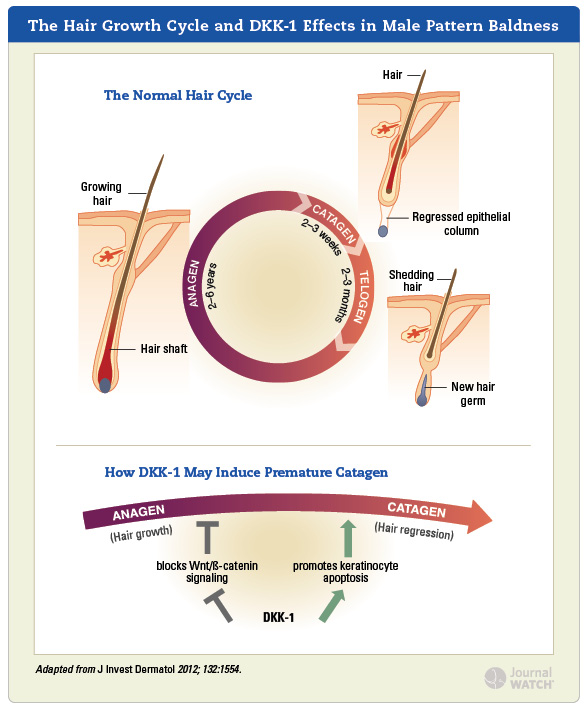
Dickkopf 1 promotes regression of hair follicles.

Kwack MH, Kim MK, Kim JC, Sung YK.
Source

Department of Immunology, School of Medicine, Kyungpook National University, Daegu, Korea.

Abstract

Recently, we suggested that Dickkopf 1 (DKK-1) is a pathogenic mediator involved in male pattern baldness. As premature catagen onset is a key characteristic of male pattern baldness, in this study, we evaluated whether DKK-1 has a role as a catagen inducer in hair cycling. Herein, we report that recombinant human DKK-1 (rhDKK-1) injection into the hypodermis of mice during anagen caused premature onset of catagen, whereas neutralizing DKK-1 antibody delayed anagen-to-catagen transition in mice. Moreover, treatment with rhDKK-1 led to a decrease in final hair follicle length, whereas DKK-1 antibody led to an increase compared with control animals. In addition, DKK-1 and DKK-1 messenger RNA expression is most upregulated in follicular keratinocytes of late anagen in depilation-induced hair cycle progression. Moreover, we observed that rhDKK-1 blocks canonical Wnt-mediated activation of β-catenin signaling and induces the proapoptotic protein Bax, resulting in apoptosis in outer root sheath keratinocytes. Taken together, our data strongly suggest that DKK-1 is involved in anagen-to-catagen transition in the hair cycle by regulating the activity of follicular keratinocytes.

Dihydrotestosterone-inducible dickkopf 1 from balding dermal papilla cells causes apoptosis in follicular keratinocytes
.

Recent studies suggest that androgen-driven alteration to the autocrine and paracrine factors produced by scalp dermal papilla (DP) cells may be a key to androgen-potentiated balding. Here, we screened dihydrotestosterone (DHT)-regulated genes in balding DP cells and found that dickkopf 1 (DKK-1) is one of the most upregulated genes. DKK-1 messenger RNA is upregulated in 3-6 hours after 50-100 nM DHT treatment and ELISA showed that DKK-1 is secreted from DP cells in response to DHT. A co-culture system using outer root sheath (ORS) keratinocytes and DP cells showed that DHT inhibits the growth of ORS cells, and neutralizing antibody against DKK-1 significantly reversed the growth inhibition of ORS cells. Analysis of co-cultured ORS cells showed a significant increment of sub-G1 apoptotic cells in response to DHT. Also, recombinant human DKK-1 inhibited the growth of ORS cells and triggered apoptotic cell death. In addition, DHT-induced epithelial cell death in cultured hair follicles was reversed by neutralizing DKK-1 antibody. Moreover, immunoblotting showed that the DKK-1 level is up in the bald scalp compared with the haired scalp of patients with androgenetic alopecia. Altogether, our data strongly suggest that DHT-inducible DKK-1 is involved in DHT-driven balding.

- - - Updated - - -

JO2012072001_zpsf2825cf1.jpg
 

squeegee

Banned
Reaction score
132
Alopecia areata though...
This is probably why D3 is working with AA which has a strong autoimmune component.

The vitamin D3 analog calcipotriol suppresses the number and antigen-presenting function of Langerhans cells in normal human skin.

Dam TN, Møller B, Hindkjaer J, Kragballe K.
Source

Department of Dermatology, University of Aarhus, Denmark.

Abstract

Local activation of T lymphocytes appears to play an important role in psoriasis and autoimmune skin disease. 1 alpha,25-dihydroxyvitamin D3 and the vitamin D3 analog calcipotriol have been shown to inhibit immune induction in vitro. The purpose of the present study was to investigate the in vivo effect of calcipotriol on Langerhans cells in normal human skin and to determine the effect of 1,25-dihydroxyvitamin D3 and calcipotriol on isolated Langerhans cells to induce autologous T-cell proliferation. Using confocal laser scanning microscopy of epidermal suction blister roofs, it was found that application of calcipotriol cream to normal human skin for 4 d resulted in a dose-dependent decrease in the number of CD1a+ cells with a dendritic morphology and in the number of dendrites per cell. The suppressive effect of calcipotriol on Langerhans cells was as strong as that of the potent corticosteroid mometasonfuroate. In Langerhans cell-enriched cell suspensions (60-97% pure) isolated from normal human skin, 1,25-dihydroxyvitamin D3 and calcipotriol (10(-8)-10(-7) M) significantly suppressed their ability to stimulate antigen-dependent T-cell proliferation. Furthermore, the vitamin D receptor was detected by Western blot analysis in the isolated Langerhans cells. Neither immunohistochemical studies nor flow cytometry of Langerhans cells showed any change in the human leukocyte antigen-DR expression after 48 h culture with antigen with or without calcipotriol. It is proposed that the inhibitory effects of the vitamin D3 on Langerhans cells may induce immunosuppression in the skin.

- - - Updated - - -


squeegee

Banned
Reaction score
132
Active D3 also significantly raises Wnt10b mRNA levels:
http://www.vitamindwiki.com/tiki-index.php?page_id=3049

and it is becoming more and more evident as far as I understand that a major cause of male pattern baldness is highly pointing to DKK1 down-regulating Wnt signaling. In addition to this and the anti-inflammatory properties of D3, I believe that topical calcitriol might be very valuable in helping DHT related male pattern baldness.

ps: love your new avatar :laugh:


This is it Princess! WNT signaling is the keyword.. and yeah active D3 should be added to the arsenal!:bigun2:

Love the dog too! This is my old avatar, just figured out how to make it work since they updated this very forum.
 

squeegee

Banned
Reaction score
132
Wnt signaling maintains the hair-inducing activity of the dermal papilla

The formation of the hair follicle and its cyclical growth, quiescence, and regeneration depend on reciprocal signaling between its epidermal and dermal components. The dermal organizing center, the dermal papilla (DP), regulates development of the epidermal follicle and is dependent on signals from the epidermis for its development and maintenance. GFP specifically expressed in DP cells of a transgenic mouse was used to purify this population and study the signals required to maintain it. We demonstrate that specific Wnts, but not Sonic hedgehog (Shh), maintain anagen-phase gene expression in vitro and hair inductive activity in a skin reconstitution assay.

http://genesdev.cshlp.org/content/14/10/1181.long

- - - Updated - - -


Keratinocyte Growth Inhibition through the Modification of Wnt Signaling by Androgen in Balding Dermal Papilla Cells

- Author Affiliations

  • Departments of Dermatology (T.K., H.T., N.K., S.K.) and Anatomy and Neurobiology (T.K., K.-I.M., M.K.), Kyoto Prefectural University of Medicine Graduate School of Medical Science, Kyoto 602-5586, Japan; and Department of Regenerative Dermatology (S.In., S.It.), Graduate School of Medicine, Osaka University, Osaka 565-0871, Japan

  • Address all correspondence and requests for reprints to: Kawata, Department of Anatomy and Neurobiology, Kyoto Prefectural University of Medicine, Kawaramachi Hirokouji, Kamigyo-ku, Kyoto 602-8566, Japan. E-mail:
Abstract

Context/Objective: Androgen induces androgenetic alopecia (Androgenetic Alopecia), which has a regressive effect on hair growth from the frontal region of the scalp. Conversely, Wnt proteins are known to positively affect mammalian hair growth. We hypothesized that androgen reduces hair growth via an interaction with the Wnt signaling system. The objective of this study was to investigate the effect of androgen on Wnt signaling in dermal papilla (DP) cells.
Design: The effect of androgen and Wnt3a on keratinocyte proliferation was measured by use of a coculture system consisting of DP cells and keratinocytes. The molecular mechanisms of androgen and Wnt pathway interactions in DP cells were examined by analyzing the expression, intracellular localization, and activity of the androgen receptor (AR) and also downstream Wnt signaling molecules.
Results: Wnt3a-dependent keratinocyte growth was suppressed by the addition of dihydrotestosterone in coculture with DP cells that were derived from Androgenetic Alopecia patients, but growth was not suppressed in coculture with DP cells from non-Androgenetic Alopecia males. Whereas DP cells from both scalp regions expressed AR protein, the expression levels of AR and cotranslocation with β-catenin, a downstream Wnt signaling molecule, were higher in DP cells of Androgenetic Alopecia patients than in DP cells from non-Androgenetic Alopecia males. In addition, significant suppression of Wnt signal-mediated transcription in response to dihydrotestosterone treatment was observed only in DP cells from Androgenetic Alopecia patients.

Conclusion: These results suggest that Wnt signaling in DP cells is regulated by androgen and this regulation plays a pivotal role in androgen’s action on hair growth.


- - - Updated - - -

A decline in tissue regenerative capacity is a hallmark of mammalian aging and is, in part, attributed to the impairment of tissue stem/progenitor cell function. It was previously shown that the age-related decline in tissue-specific progenitor cell activity is modulated by factors that are present in the serum.[SUP]58[/SUP] In line with this study, Brack et al[SUP]52[/SUP] provided evidence showing that systemic factors in the serum of aged mice activate canonical Wnt signaling and contribute to age-related decline in skeletal muscle regeneration. The authors showed that skeletal muscle stem cells (satellite cells) convert from a myogenic to a fibrogenic lineage when exposed to aged serum and that canonical Wnt signaling is enhanced in skeletal muscle of aged mice and in cultured satellite cells exposed to aged serum. Moreover, skeletal muscle regeneration in young animals was attenuated by Wnt3A treatment, whereas impaired muscle regeneration in aged mice was restored by inhibition of canonical Wnt signaling. These observations suggest that activation of Wnt signaling by the “Wnt-like substance” present in the serum of aged organisms contributes to a decline in tissue stem cell function and impaired tissue regeneration associated with aging.

http://circres.ahajournals.org/content/107/11/1295.full

- - - Updated - - -

Oxidative Stress Antagonizes Wnt Signaling in Osteoblast
Precursors by Diverting

-Catenin from T Cell Factor- to
Forkhead Box O-mediated Transcription

http://www.jbc.org/content/282/37/27298.full.pdf


- - - Updated - - -

I much prefer the other one. For some reason I can't take seriously what you post with that old avatar.

Why so serious? Because Kermit the frog was better?:p You will get over it Odal! LOL

- - - Updated - - -

[h=1]The vitamin D receptor, the skin and stem cells.[/h]Luderer HF, Demay MB.
[h=3]Source[/h]Endocrine Unit, Massachusetts General Hospital, Harvard Medical School, 50 Blossom St, Thier 11, Boston, MA 02114, USA.

[h=3]Abstract[/h]The active metabolite of vitamin D, 1,25-dihydroxyvitamin D, has been shown to have pro-differentiation and antiproliferative effects on keratinocytes that are mediated by interactions with its nuclear receptor. Other cutaneous actions of the vitamin D receptor have been brought to light by the cutaneous phenotype of humans and mice with non-functional vitamin D receptors. Although mice lacking functional vitamin D receptors develop a normal first coat of hair, they exhibit impaired cyclic regeneration of hair follicles that leads to the development of alopecia. Normal hair cycling involves reciprocal interactions between the dermal papilla and the epidermal keratinocyte. Studies in mice with targeted ablation of the vitamin D receptor demonstrate that the abnormality in the hair cycle is due to a defect in the keratinocyte component of the hair follicle. Furthermore, expression of mutant vitamin D receptor transgenes in the keratinocytes of vitamin D receptor knockout mice demonstrates that the effects of the receptor that maintain hair follicle homeostasis are ligand-independent. Absence of a functional vitamin D receptor leads to impaired function of keratinocyte stem cells, both in vivo and in vitro. This is manifested by impaired cyclic regeneration of the hair follicle, a decrease in bulge keratinocyte stem cells with ageing and an abnormality in lineage progression of these cells, leading to their preferential differentiation into sebocytes.
Copyright (c) 2010. Published by Elsevier Ltd.

- - - Updated - - -

[h=1]Vitamin D receptor is essential for normal keratinocyte stem cell function[/h]
[h=2]Abstract[/h] The major physiological role of the vitamin D receptor (VDR) is the maintenance of mineral ion homeostasis. Mutation of the VDR, in humans and mice, results in alopecia. Unlike the effects of the VDR on mineral ion homeostasis, the actions of the VDR that prevent alopecia are ligand-independent. Although absence of the VDR does not prevent the development of a keratinocyte stem cell niche in the bulge region of the hair follicle, it results in an inability of these stem cells to regenerate the lower portion of the hair follicle in vivo and impairs keratinocyte stem cell colony formation in vitro. VDR ablation is associated with a gradual decrease in keratinocyte stem cells, accompanied by an increase in sebaceous activity, a phenotype analogous to that seen with impaired canonical Wnt signaling. Transient gene expression assays demonstrate that the cooperative transcriptional effects of β-catenin and Lef1 are abolished in keratinocytes isolated from VDR-null mice, revealing a role for the unliganded VDR in canonical Wnt signaling. Thus, absence of the VDR impairs canonical Wnt signaling in keratinocytes and leads to the development of alopecia due to a defect in keratinocyte stem cells.

http://www.pnas.org/content/104/22/9428.full

- - - Updated - - -

You're not comparing it to Proctor's site..are you? :woot:

I don't know of anyone else that does custom products for individuals. Or even small batches. At least not liposomal-type topicals.

He's always ended up responding to my emails..eventually. But I feel your frustration..this is the longest it's been for me too :(

Is Elsom cGMP qualified? I am afraid that the FDA gonna shut it down..
 

Jacob

Senior Member
Reaction score
44
The one thing I just hate about this forum... If someone edits(Updates) their post the thread doesn't show as having a new post. I'm referring to when someone replies in a thread that they last posted in...their latest "post" just gets morphed into their previous one. While I can see why that's done..it's cool and all..it really prevents ppl from seeing such posts.

Of course if someone just edits their last post..then it's their own fault for sure that no one saw it :p


So I just happened to come across your addition there squeegee. I'll ask him....
 

squeegee

Banned
Reaction score
132
The one thing I just hate about this forum... If someone edits(Updates) their post the thread doesn't show as having a new post. I'm referring to when someone replies in a thread that they last posted in...their latest "post" just gets morphed into their previous one. While I can see why that's done..it's cool and all..it really prevents ppl from seeing such posts.

Of course if someone just edits their last post..then it's their own fault for sure that no one saw it :p


So I just happened to come across your addition there squeegee. I'll ask him....

LOL I know.. there is nothing to separate the crap when a member post several times in a row.. Pain in the arse! Jacob you will probably get an answer by next week or so lol I got my second e-mail answered couple days ago.. the guy seems to be very knowledgeable but awful when it comes to communication!

- - - Updated - - -

Hairloss is just a big mitochondrial dysfunction.. all the symptoms are there: Glutathione depletion, lipid oxidation.. Vitamin D3 receptors are damaged by Oxidative stress and chronic inflammation.. This is why you hair don't grow back. Osteoporosis run on the same pattern but internally. When a balding dude add topical l-carnitine ..hair are growing back..restoring mitochondria energy production..when a balding dude add minoxidil.. the hair are growing back.. why.. NO control Mitochondrial function.. simple as that.
 

Sparky4444

Senior Member
Reaction score
44
ok boys..can we review here??? ..I got lost along the way somehow...Are we talking topical Vitamin D and topical Glutathione??

The initial post is about hair regrowth on a child using topical Vitamin D...a different form of Vitamin D you can't buy over the counter...Now, you would think with this kind of regrowth - granted it isn't male pattern baldness, I know, but regardless -- you would expect this to be tried on someone with common male pattern baldness...but so far, haven't heard of it yet...
 

squeegee

Banned
Reaction score
132
úp cho ngÃ*y cu?i tu?n nÃ*o :D


GOOGLE TRANSLATION: Ã º p for old Russia * y? I from? N na * o wtf? spammer are taking over the ****ing web.. **** them!
 

squeegee

Banned
Reaction score
132
I am trying it, seeing small pigmented (not vellus, but very dark hair) in a 2 square inch area that was slick bald for years, and i mean slick bald, just buzzed head completely today as i am using the cream and it is difficult to apply when hair gets longer, (scalp solution seems always out of stock in that one online pharmacy), wife told me last night "you hair is getting very dark" and that made me happy :woot: [she is the actual princessRambo, her handle, when we play online games, i always thought it hilarious when she kills someone and it says: "you were killed by princessRambo" :uglylol:] anyway, i think there is something to this, it has been 6 weeks so far, i will keep you posted in a couple months with pictures. I should note that I also take 3000 ml omega 3 (not fish oil amount, but actual quantity of omega 3 ingested), 6gram of evening primrose oil daily and multi vits, but i have been doing that for a while, not sure if that has interfered with the results i am seeing, i thought i would let you guys know that, as a full disclosure. Anyhow, here is a research I found noteworthy: http://www.ncbi.nlm.nih.gov/pubmed/15538745


[FONT=.HelveticaNeueUI]http://www.fertstert.org/article/S0015-0282(11)02791-9/abstract[/FONT]

http://informahealthcare.com/doi/abs/10.3109/09513590.2012.683079

This is also an interesting video, please note how he talks about injecting vd3 around 12mn into the video. I think topical cream/solution of calcipotriol can actually reach the derma papilla cell. I am thinking derma roller + calcipotriol, would that work better without inducing hypercalcemia ? :doh:

Really good post Princess! Keep us updated with the results!
 

IDW2BB

Established Member
Reaction score
19
http://www.ncbi.nlm.nih.gov/pmc/articles/PMC3875938/


from the link:

Although 1,25(OH)2D3 inhibits β-catenin/TCF transcriptional activity in colon and other cancer cells, the upregulation of the Wnt/β-catenin pathway by either ligand-activated or unliganded VDR has been described in osteoblasts and keratinocytes, where it promotes bone formation and hair follicle differentiation, respectively. Thus, the interplay between Wnt/β-catenin pathway and 1,25(OH)2D3/VDR seems to depend on the cell or tissue type.

Wnt signaling promotes the differentiation of bone marrow-derived mesenchymal stem cells to bone while it represses their differentiation to other cell types, such as adipocytes [110,111]. Some 1,25(OH)2D3 effects in bone are similar to those of Wnt, suggesting a crosstalk between both pathways. Indeed, 1,25(OH)2D3 induces the expression of the Wnt co-receptor Lrp5 in mouse osteoblasts [112,113], while represses that of the Wnt inhibitors Dkk-1 and Sfrp2 in mouse bone marrow-derived mesenchymal stem cells [114]. These effects support a role of 1,25(OH)2D3 stimulating Wnt signaling in normal bone. However, the interplay between these two pathways seems to change in tumor tissue, as it has been reported that 1,25(OH)2D3 inhibits Wnt/β-catenin signaling in SaOS2 osteosarcoma cells [102].

Ligand-independent actions of VDR on Wnt canonical signaling have also been reported. In the skin, absence of Vdr, but not of 1,25(OH)2D3, results in alopecia in mice [115], and two independent groups have demonstrated that this is due, at least in part, to impaired Wnt/β-catenin signaling in keratinocytes [116,117]. In this regard, Pálmer et al. have shown that VDR is a Wnt effector and that β-catenin behaves as a VDR co-activator in the skin to induce transcription of genes associated with differentiation of hair follicle lineages [117]. Although this effect is largely 1,25(OH)2D3-independent, it is enhanced by the hormone. More recently, Luderer et al. have reported a direct interaction between VDR and LEF1 that is independent of both ligand and β-catenin, and that is required for normal canonical Wnt signaling in keratinocytes [118]. Interestingly, Lef1 knock-out mice develop alopecia at an early age [119] and transgenic mice expressing a dominant negative Lef1 in keratinocytes also show a phenotype that resembles that of Vdr−/− mice [120]. Therefore, although the in vivo significance of the VDR-LEF1 interaction is not yet clear, it may contribute to at least some of the ligand-independent effects of VDR in the skin. Similarly to what happens in bone, this interplay seems to change in the tumoral context. The development of trichofolliculomas (benign hair follicle tumors) induced by prolonged activation of β-catenin in the skin is inhibited by the 1,25(OH)2D3 analogue EB1089 [117]. In addition, undifferentiated tumors resembling basal cell carcinomas instead of trichofolliculomas are developed by β-catenin activation in Vdr−/− mice [117]. Accordingly, keratinocytes lacking VDR present decreased E-cadherin expression, increased β-catenin/TCF transcriptional activity, and a higher proliferation rate; whereas VDR overexpression or 1,25(OH)2D3 treatment has the opposite effect [121,122]. These results suggest that 1,25(OH)2D3/VDR suppresses epidermal tumor formation by limiting the hyperproliferative actions of β-catenin in the skin. Surprisingly, epidermal β-catenin ablation cannot reduce the increased number of UVB-induced tumors developed by epidermis-specific Vdr knock-out mice, suggesting that VDR has β-catenin-independent anticancer functions in this model [122].
 
Top