waynakyo
Experienced Member
- Reaction score
- 466
Maybe was posted/discussed before:
http://onlinelibrary.wiley.com/doi/10.1111/bjd.12921/abstract
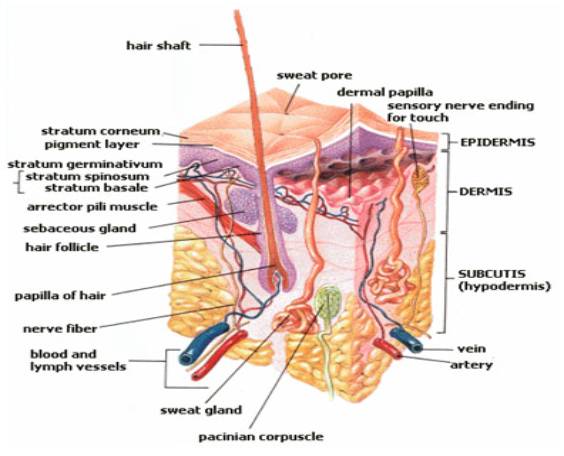
[h=4][FONT=Verdana, Arial, Tahoma, Calibri, Geneva, sans-serif]Background[/FONT][/h][FONT=Verdana, Arial, Tahoma, Calibri, Geneva, sans-serif]Androgenic alopecia (Androgenetic Alopecia) is the most common hair loss condition in men and women. Hair loss is caused by follicle miniaturization, which is largely irreversible beyond a certain degree of follicular regression. In contrast, hair loss in telogen effluvium (Telogen Effluvium) is readily reversible. The arrector pili muscle (APM) connects the follicle to the surrounding skin.[/FONT]
[h=4][FONT=Verdana, Arial, Tahoma, Calibri, Geneva, sans-serif]Objectives[/FONT][/h][FONT=Verdana, Arial, Tahoma, Calibri, Geneva, sans-serif]To compare histopathological features of the APM in Androgenetic Alopecia and Telogen Effluvium.[/FONT]
[h=4][FONT=Verdana, Arial, Tahoma, Calibri, Geneva, sans-serif]Methods[/FONT][/h][FONT=Verdana, Arial, Tahoma, Calibri, Geneva, sans-serif]Archival blocks of 4-mm scalp punch biopsies from eight patients with Androgenetic Alopecia and five with Telogen Effluvium were obtained. New 4-mm biopsies from five normal cases were used as controls. Serial 7-μm sections were stained with a modified Masson's trichrome stain. ‘Reconstruct’ software was used to construct and evaluate three-dimensional images of the follicle and APM.[/FONT]
[h=4][FONT=Verdana, Arial, Tahoma, Calibri, Geneva, sans-serif]Results[/FONT][/h][FONT=Verdana, Arial, Tahoma, Calibri, Geneva, sans-serif]The APM degenerated and was replaced by adipose tissue in all Androgenetic Alopecia specimens. Remnants of the APM remained attached to the hair follicle. There was no fat in the normal skin specimens. Fat was seen in two of five Telogen Effluvium specimens but could be attributed to these patients also showing evidence of Androgenetic Alopecia. Quantitative analysis showed that muscle volume decreased and fat volume increased significantly (P < 0·05) in Androgenetic Alopecia compared with controls.[/FONT]
[h=4][FONT=Verdana, Arial, Tahoma, Calibri, Geneva, sans-serif]Conclusions[/FONT][/h][FONT=Verdana, Arial, Tahoma, Calibri, Geneva, sans-serif]APM degeneration and replacement with fat in Androgenetic Alopecia has not previously been described. The underlying mechanism remains to be determined. However, we speculate that this phenomenon might be related to depletion of stem or progenitor cells from the follicle mesenchyme, explaining why Androgenetic Alopecia is treatment resistant.[/FONT]
http://onlinelibrary.wiley.com/doi/10.1111/bjd.12921/abstract
[h=4][FONT=Verdana, Arial, Tahoma, Calibri, Geneva, sans-serif]Background[/FONT][/h][FONT=Verdana, Arial, Tahoma, Calibri, Geneva, sans-serif]Androgenic alopecia (Androgenetic Alopecia) is the most common hair loss condition in men and women. Hair loss is caused by follicle miniaturization, which is largely irreversible beyond a certain degree of follicular regression. In contrast, hair loss in telogen effluvium (Telogen Effluvium) is readily reversible. The arrector pili muscle (APM) connects the follicle to the surrounding skin.[/FONT]
[h=4][FONT=Verdana, Arial, Tahoma, Calibri, Geneva, sans-serif]Objectives[/FONT][/h][FONT=Verdana, Arial, Tahoma, Calibri, Geneva, sans-serif]To compare histopathological features of the APM in Androgenetic Alopecia and Telogen Effluvium.[/FONT]
[h=4][FONT=Verdana, Arial, Tahoma, Calibri, Geneva, sans-serif]Methods[/FONT][/h][FONT=Verdana, Arial, Tahoma, Calibri, Geneva, sans-serif]Archival blocks of 4-mm scalp punch biopsies from eight patients with Androgenetic Alopecia and five with Telogen Effluvium were obtained. New 4-mm biopsies from five normal cases were used as controls. Serial 7-μm sections were stained with a modified Masson's trichrome stain. ‘Reconstruct’ software was used to construct and evaluate three-dimensional images of the follicle and APM.[/FONT]
[h=4][FONT=Verdana, Arial, Tahoma, Calibri, Geneva, sans-serif]Results[/FONT][/h][FONT=Verdana, Arial, Tahoma, Calibri, Geneva, sans-serif]The APM degenerated and was replaced by adipose tissue in all Androgenetic Alopecia specimens. Remnants of the APM remained attached to the hair follicle. There was no fat in the normal skin specimens. Fat was seen in two of five Telogen Effluvium specimens but could be attributed to these patients also showing evidence of Androgenetic Alopecia. Quantitative analysis showed that muscle volume decreased and fat volume increased significantly (P < 0·05) in Androgenetic Alopecia compared with controls.[/FONT]
[h=4][FONT=Verdana, Arial, Tahoma, Calibri, Geneva, sans-serif]Conclusions[/FONT][/h][FONT=Verdana, Arial, Tahoma, Calibri, Geneva, sans-serif]APM degeneration and replacement with fat in Androgenetic Alopecia has not previously been described. The underlying mechanism remains to be determined. However, we speculate that this phenomenon might be related to depletion of stem or progenitor cells from the follicle mesenchyme, explaining why Androgenetic Alopecia is treatment resistant.[/FONT]